About diabetic eye screening - Your diabetic eye screening results
You will receive your results in the post within eight weeks of your appointment. Your doctor and diabetes specialist (if you have one) will also receive a copy.
Contact us if you have not received your results within eight weeks or have any questions about the results.
The letter you receive will tell you if you have diabetic retinopathy or not and what happens next.
Most people will not have diabetic retinopathy and will be invited for their next screening appointment when due.
Some people will be asked to have another test. This will be because we were unable to get a result from the photographs. This is not unusual.
Your results letter will tell you if you have diabetic retinopathy. It will tell you if you need further tests or treatment. This may be with a specialist optometrist or at a hospital. They will send you an appointment if you need further tests or treatment.
Your results explained
Each eye is assessed for a retinopathy and maculopathy grade.
Retinopathy is damage to the blood vessels supplying the back of the eye (the retina). We grade retinopathy with an R grade from 0 to 3.
Maculopathy – Sometimes we see something in a specific area of the back of your eye called the macula. We grade maculopathy with an M grade from 0 to 1.
The result letter will not include the grade of retinopathy that we have found. More detailed information is shared with your doctor and diabetic specialist (if you have one).
Contact your doctor or us if you would like to receive a more detailed summary of your results.
The type of results we provide are as follows:
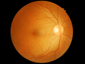
No diabetic retinopathy – classified as R0M0
If we do not find any retinopathy in your eyes, we will send you a letter telling you that your photographs show no retinopathy.
This image is an example of an eye with no retinopathy:
You will be invited for your screening appointment when due. It is important to attend your appointment when invited.
Background (some) diabetic retinopathy changes – classified as R1
We will tell you if we have seen background (some) retinopathy in your photographs. This does not need further investigation. Finding these changes does not mean that retinopathy will continue to get worse. Sometimes the changes will appear and then go away again.
You will be invited for your screening appointment when due. It is important to attend your appointment when invited.
It is important to keep healthy, by managing and treating your diabetes Visit Diabetes management | taking care of your diabetes | Diabetes UK or talk to your health professional for more information.
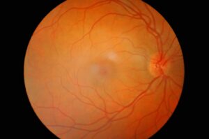
The image below is an example of background retinopathy changes. If you look closely there are very small red dots showing some leaking blood vessels.
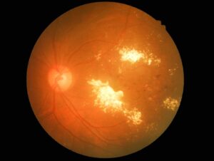
Referable retinopathy changes – classified as R2 or R3
Your letter will tell you if you have retinopathy. This is called pre-proliferative (R2) or proliferative (R3) retinopathy and you should be seen by an eye specialist. In some cases, treatment for diabetic retinopathy is needed.
It is important to attend any further appointments. Any changes can then be monitored and referred for treatment at a hospital if required.
Contact your doctor if you are worried or have any questions.
This is an example of an R2 image, showing leaking blood vessels leaking into the eye:
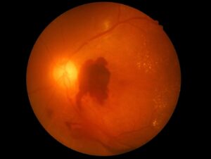
This is an example of an R3 image. This shows a large bleed within the eye:
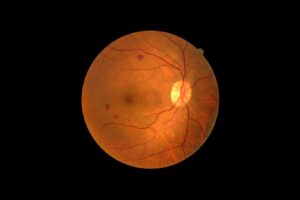
Maculopathy – classified as M1
If we see maculopathy we will ask a specialist to carry out a more detailed check of your eyes.
The image below is an example of an eye with maculopathy (M1):
What happens if I am called back for further tests?
Depending on the results of your screening, you may be asked to have screening appointments more often. We may also refer you to an eye specialist for further assessment and treatment. The eye specialist will contact you with details of your appointment.
Contact the clinic if you need further information.
What happens next?
If your photographs were not clear enough to give a result, you will be offered another appointment within three months.
If you are being followed-up at a hospital eye clinic, you will not be invited for screening until you have been discharged by an eye specialist.
If you are pregnant, you will be offered screening more often during your pregnancy.
If you are not able to take part in diabetic eye screening, contact your local Optician who may be able to carry out home visits. Some Optometrists may provide a free visit, depending on your circumstances.
Contact your optician or doctor if you notice any changes in your eyesight. Do not wait for your next screening appointment.
Page last reviewed: 24th March 2026